
At Baseside Dental Office, we have invested in a core set of diagnostic and treatment tools that we use consistently across our practice. Our equipment choices reflect a specific clinical philosophy: accurate information leads to better decisions, and better decisions lead to better outcomes.
Below is an overview of the key equipment in our practice and how each piece contributes to your care.
Dental CT (Cone Beam Computed Tomography)
Seeing What Standard X-Rays Cannot Show
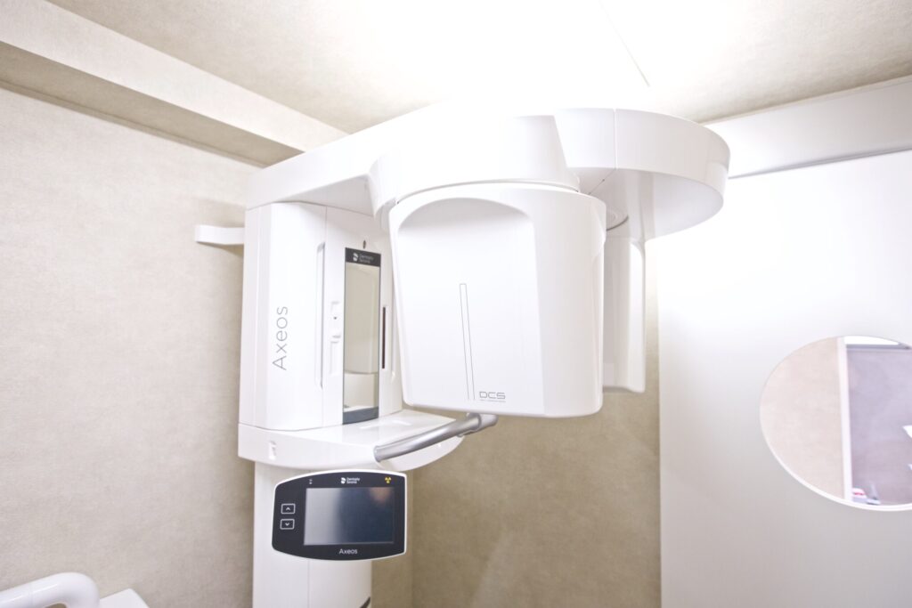
A dental CT — also called a CBCT (cone beam computed tomography) scanner — produces a three-dimensional image of the jaw, teeth, bone, joints, and surrounding structures. Unlike conventional flat X-rays, which compress a three-dimensional anatomy into a two-dimensional image, CT data allows us to examine any cross-section of the mouth from any angle.
At our practice, CT is used routinely at new patient exams and for any case where three-dimensional information is clinically meaningful. This includes:
Implant planning — evaluating bone volume, density, and the precise location of nerves and sinuses before placing any implant or fabricating a surgical guide.
Airway screening — measuring the volume and cross-sectional shape of the upper airway to identify narrowing that may be associated with obstructive breathing during sleep.
Endodontic assessment — identifying canal anatomy, periapical pathology, and root fractures that are not detectable on standard X-rays.
TMJ evaluation — assessing condylar position, joint space, and bony changes to the temporomandibular joint in three dimensions.
Periodontal assessment — evaluating the level and pattern of alveolar bone support around teeth with greater precision than is possible with periapical X-rays alone.
We show you your CT images at your appointment. Understanding what your own anatomy looks like is part of the informed consent process — not an add-on.
Intraoral Scanner
Precise Digital Impressions
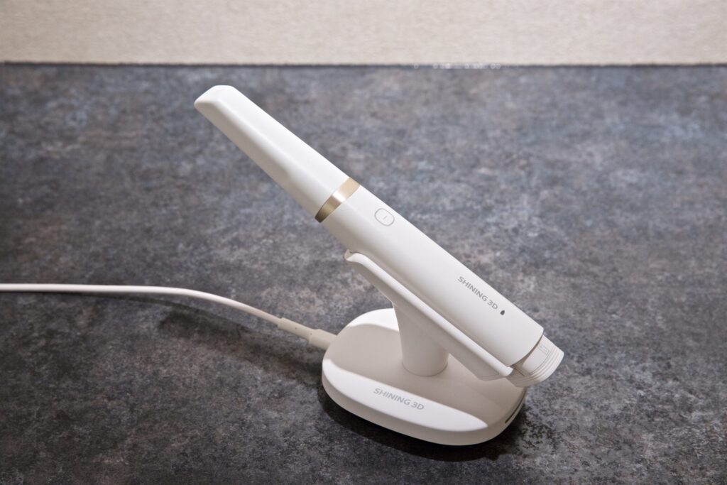
The intraoral scanner is an optical scanner that creates a precise three-dimensional digital model of your teeth and surrounding soft tissue in real time. It replaces the traditional impression process — no trays, no impression material, no extended periods of holding your mouth open with a mouthful of putty.
The scan takes only a few minutes per arch and produces a highly accurate digital record that can be used for:
- Crown, bridge, and veneer fabrication
- Implant and surgical guide planning
- Night guard and deprogrammer fabrication
- Invisalign aligner treatment planning
- Documenting the baseline position of your teeth for future comparison
- Visualizing your current bite relationship on-screen during your appointment
Patients who have had uncomfortable experiences with traditional impressions — particularly those with a strong gag reflex — often find the scanning process significantly easier to tolerate.
Dental Operating Microscope
Magnification That Changes What’s Possible
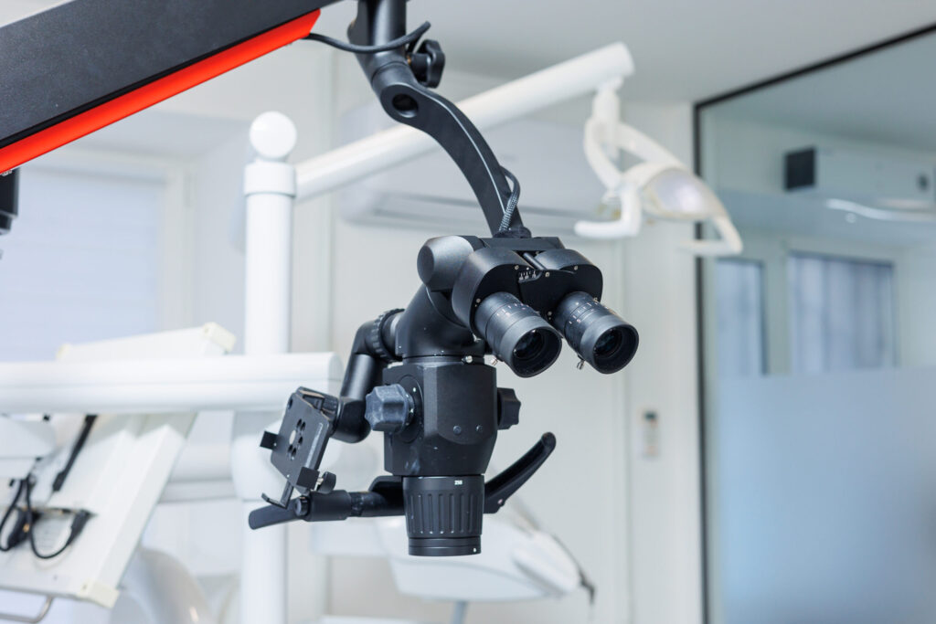
A dental operating microscope — sometimes called a surgical microscope or DOM — provides illuminated, magnified visualization of the treatment area at up to several dozen times normal size. The clinical difference between working with and without a microscope is not simply one of comfort or preference — it is a difference in what can be seen, and therefore what can be done.
We use the microscope for:
Root canal treatment — identifying all canal orifices, tracking the canal path through curved anatomy, detecting isthmuses and accessory canals, and confirming complete debridement before obturation.
Crown and veneer margin assessment — evaluating the precision of preparation margins and the fit of restorations at a level of detail that is not possible without magnification.
Crack detection — identifying cracks in tooth structure that would be invisible to the naked eye and may explain unexplained pain or recurrent restoration failure.
Periodontal and surgical procedures — improving precision during soft tissue procedures and surgical interventions.
The microscope does not replace clinical judgment — but it significantly expands the information available to inform it.
EMS Airflow Prophylaxis Master
Evidence-Based Biofilm Removal — Comfortable, Thorough, Gentle
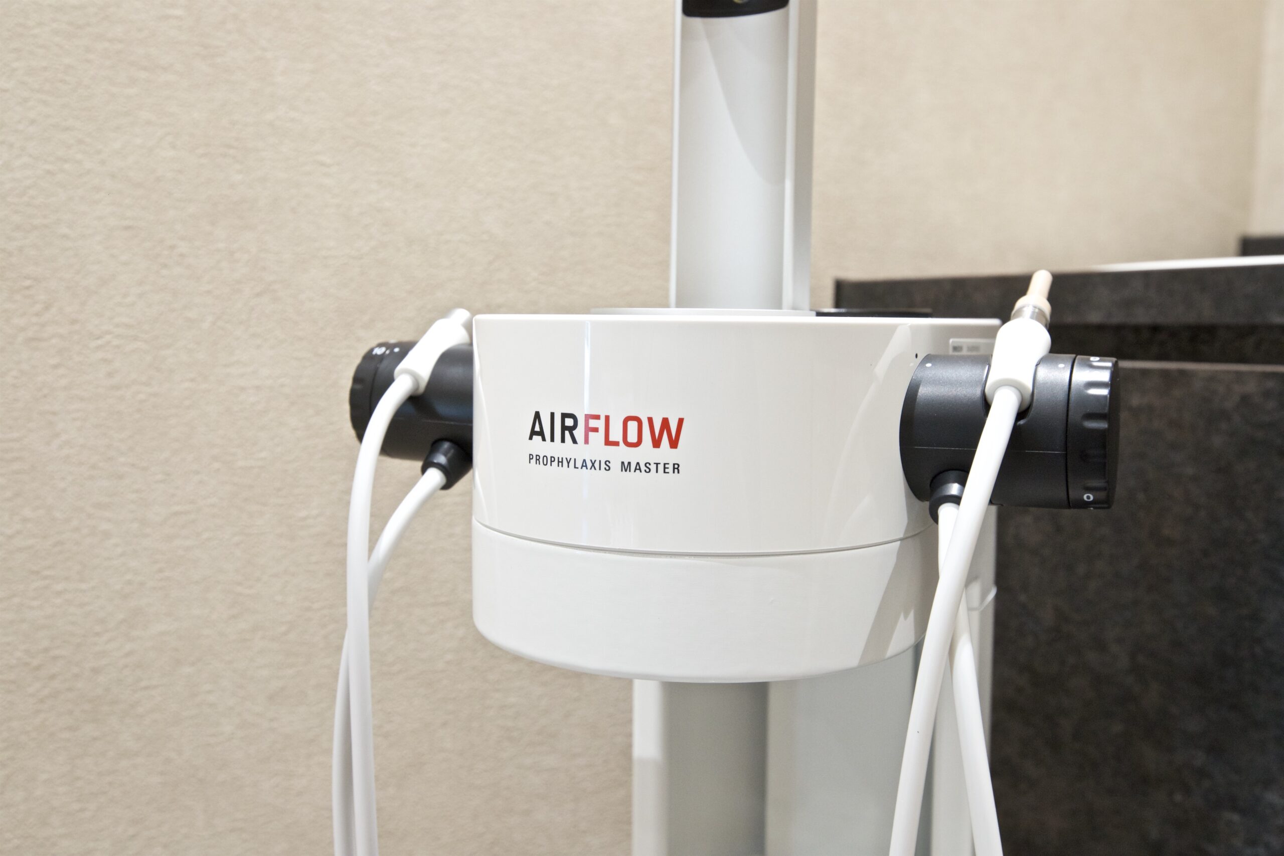
The EMS Airflow Prophylaxis Master is the delivery system for Guided Biofilm Therapy (GBT) — a structured, evidence-based protocol for professional oral biofilm management. Rather than relying primarily on manual scaling with hand instruments, GBT uses the Airflow device to disrupt and remove biofilm using a controlled stream of warm water, air, and a fine powder.
The clinical advantages of this approach include:
Reduced surface trauma — the Airflow powder is gentler on enamel and root surfaces than metal scalers, which is particularly relevant for patients with exposed root surfaces or areas of enamel hypomineralization.
Subgingival capability — the Perioflow nozzle attachment allows biofilm removal from within the periodontal pocket up to 4mm without the mechanical trauma associated with traditional subgingival scaling.
Comfort — most patients who have experienced both traditional scaling and Airflow GBT prefer the latter. The warm water temperature and reduced mechanical contact contribute to a noticeably more comfortable experience.
Thoroughness — the biofilm disclosure step at the start of the GBT protocol makes visible exactly where biofilm has accumulated, ensuring that cleaning is systematic rather than intuitive.
We use this system for all professional cleaning appointments. It forms the foundation of our preventive maintenance program and is used in conjunction with traditional hand and ultrasonic scalers when calculus removal is indicated.